Department Of Pulmonology
The Expert Team of pulmonologists is highly specialized in complex surgical and medical procedures and is experienced in treating the patients of all ages with utmost care and as per the complete satisfaction of the individuals. The pulmonologists at BIMS hospital work in collaboration with the experts of almost all other specialities in order to deliver personalised and effective care to the patient.
OVERVIEW:
Pulmonology is an area of medicine that focuses on the health of the respiratory system. Pulmonologists treat everything from asthma to tuberculosis.
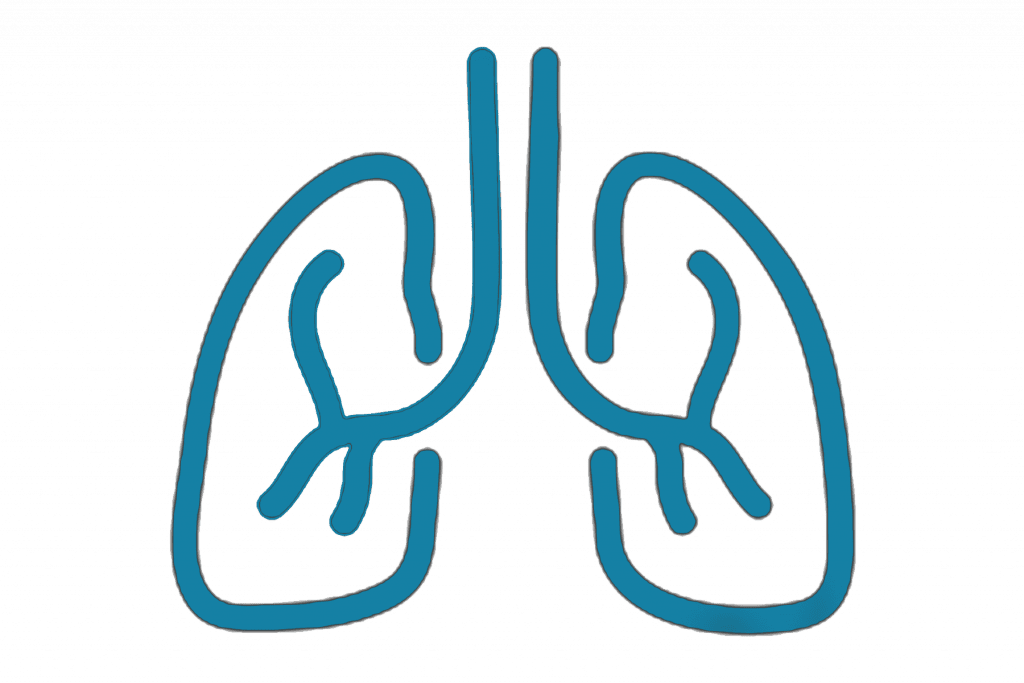
WHAT IS THE RESPIRATORY SYSTEM?
The respiratory system includes the organs that help you breathe. The three major parts of this system are the airway, the lungs, and the respiratory muscles.
The Airway Includes the:
- Nose
- Mouth
- Pharynx
- Larynx
- Trachea
- Bronchi
- Bronchioles
- Alveoli
You use several muscles during respiration. The most notable is the diaphragm. The other muscles are categorized in groups, including:
- Intercostals muscles, which help with inhalation
- Accessory muscles, which help inhalation but don’t play a primary role
- Exhalation muscles, which help with forceful or active exhalation.
WHAT IS PULMONOLOGIST?
These specialists diagnose and treat conditions that affect the respiratory system in men and women, as well as children. Pulmonologists have expertise in the following types of respiratory disorders:
- Infectious
- Structural
- Inflammatory
- Neo plastic, which means having to do with a tumour
- Autoimmune
In some instances, this extends to the cardiovascular system. Certain conditions, such as pulmonary vascular disease, can first affect the respiratory system but go on to affect other organs in the body.
WHAT IS PULMONOLOGY?
Pulmonology is a field of medicine that focuses specifically on diagnosing and treating disorders of the respiratory system.
Subspecialties of Pulmonology Include:
- Interstitial lung disease, which focuses on lung diseases marked by persistent inflammation and scarring
- Interventional pulmonology, which employs multidisciplinary care to treat airway disorders, lung cancer, and pleural diseases.
- Lung transplantation, management before and after surgery
- Neuromuscular disease, which refers to conditions that occur due to respiratory muscle failure.
- Obstructive lung disease, which involves airway narrowing or obstruction.
- Sleep-disordered breathing
WHAT CONDITIONS DO PULMONOLOGISTS TREAT?
Conditions pulmonologists commonly treat include:
- Asthma
- Bronchiectasis, a condition that involves inflammation and excess mucus
- Bronchitis, which happens when you have inflamed lower airways
- Chronic obstructive pulmonary disease (COPD), which causes an airflow blockage
- Emphysema, which happens when the alveoli in your lungs are damaged
- Interstitial lung disease, which affect the space and tissue within the lung
- Occupational Lung Disease, which can occur due to the inhalation of dusts, chemicals, or proteins
- Obstructive sleep apnea, which causes your breathing to slow or stop entirely when you’re sleeping.
- Lung infection by pneumonia virus, bacteria, fungi, parasites.
- Pleural Disease: Pleural effusion (Fluid collection in pleura), pnemno thorax (Air collection) in pleura
- Hemothorax
- Pulmonary Embolism – Blood clot in pulmonary vessels.
- Pulmonary artery Hypertension.
WHAT PROCEDURES DO PULMONOLOGISTS USE?
Pulmonologists can use and interpret exams and tests to help determine a lung-related diagnosis. These may include the following:
- CT scan to get detailed images of the bones, muscles, fat organs, and blood vessels in your chest.
- Chest Fluoroscopy, an X-ray test to see how well your lungs are function.
- Chest ultrasound to examine the organs and other chest structures
- Pleural biopsy to remove a small tissue sample from the pleura, which is the membrane that surrounds your lungs.
- Pulmonary function test, a breathing test to see how well your lungs are working.
- Pulse oximetry test to determine the oxygen saturation level in your blood.
- Thoracentesis to remove and sample fluid from around your lungs
- Chest tube to remove air or fluid from around your lungs.
- Bronchoscopy to examine your airway and determine if you have any issues in your trachea, lower airways, throat, or larynx.
- Sleep study to help diagnose sleep disorders, such as sleep apnea.
In the case of more serious lung diseases and conditions, a pulmonologist may refer you to a chest surgeon for procedures, such as a lobectomy to remove a portion of a diseased lung or a lung transplant.
WHEN SHOULD YOU SEE A PULMONOLOGIST?
If you are having any unusual symptoms, you should meet with your primary care doctor. They will perform a medical exam and assess your overall condition. They may refer you to a pulmonologist if you:
- Have difficulty breathing
- Have a persistent cough
- Regularly cough up blood or mucus
- Smoke
- Have unexplained weight loss
- Have trouble exercising due to breathing problems.
WHAT KINDS OF PROCEDURE PULMONOLOGISTS DO?
Pulmonologists can do special procedures such as:
- Pulmonary hygiene: This clears fluid and mucus from your lungs.
- Airways ablation: This opens blocked air passages or eases difficult breathing.
- Biopsy: This takes tissue samples to diagnose disease.
- Bronchoscopy: This looks inside your lungs and airways to diagnose disease.
CHRONIC OBSTRUCTIVE PULMONARY DISEASE (COPD):
Chronic obstructive pulmonary disease, commonly referred to as COPD, is a group of progressive lung diseases.
The most common of these diseases are emphysema and chronic bronchitis. Many People with COPD have both of these COPD have both of these conditions.
Emphysema slowly destroys air sacs in your lungs, which interferes with outward air flow. Bronchitis causes inflammation and narrowing of the bronchial tubes, which allows mucus to build up.
WHAT ARE THE SYMPTOMS OF CHRONIC OBSTRUCTIVE PULMONARY DISEASE (COPD)?
COPD makes it harder to breath. Symptoms may be mild at first, beginning with intermittent coughing and shortness of breath. As it progresses, symptoms can become more constant to where it can become increasingly difficult to breath even on resting condition.
You may experience wheezing and tightness in the chest or have excess sputum production. Some people with COPD have acute exacerbations, which are flare-ups of severe symptoms.
EARLY SYMPTOMS:
At first, symptoms of COPD can be quite mild. You might mistake them for a cold.
Early Symptoms include:
- Occasional shortness of breath, especially after exercise
- Mild but recurrent cough
- Needing to clear your throat often, especially first thing in the morning.
You might start making subtle changes, such as avoiding stairs and skipping physical activities.
WORSENING SYMPTOMS:
Symptoms can get progressively worse and harder to ignore. As the lungs become more damaged, you may experience:
- Shortness of breath, after even mild forms of exercise like walking up a flight of stairs.
- Wheezing, which is a type of higher – pitched noisy breathing, especially during exhalations
- Chest tightness
- Chronic cough, with or without mucus
- Need to clear mucus from your lungs everyday
- Frequent colds, flu, or other respiratory infections
- Lack of energy
In later stages of COPD, symptoms may also include:
- Fatigue
- Swelling of the feet, ankles, or legs
- Weight loss
WHAT CAUSES COPD?
Most people with COPD are at least 40 years old and have at least some history of smoking. The longer and more tobacco products you smoke, the greater your risk of COPD is.
In addition to cigarette smoke, cigar smoke, pipe smoke, and second hand smoke can cause COPD. Your rist of COPD is even greater if you have asthma and smoke.
OTHER CAUSES:
You can also develop COPD if you are exposed to chemicals and fumes in the workplace. Long-Term exposure to air pollution and inhaling dust can also cause COPD.
In developing countries, along with tobacco smoke, homes are often poorly ventilated, forcing families to breath fumes from burning fuel used for cooking and heating.
There may be a genetic predisposition to developing COPD. Up to an estimated 5% of people with COPD have a deficiency in a protein called alpha-1-antitypsin.
This deficiency causes the lungs to deteriorate and also can affect the liver. There may be other associated genetic factors at play as well.
DIAGNOSING COPD:
Diagnosis is based on symptoms, a physical exam, and diagnostic test results. When you visit the doctor, be sure to mention all of your symptoms. Tell your doctor if:
- You are smoker or have smoked in the past
- You are exposed to lung irritants on the job
- You are exposed to a lot of second hand smoke
- You have a family history of COPD
- You have asthma or other respiratory conditions
- You take over-the-counter or prescription medications.
EXAM AND TESTS:
During the physical exam, your doctor will use a stethoscope to listen to your lungs as you breathe. Based on all this information, your doctor may order some of these tests to get a more complete picture:
- Spirometry is non-invasive test to assess lung function. During the test, you will take a deep breath and then blow into a tube connected to the spirometer.
- Imaging Tests, like a chest X-ray or CT scan. These images can provide a detailed look at your lungs, blood vessels, and heart.
- An arterial blood gas test. This involves taking a blood sample from an artery to measure your blood oxygen, carbon dioxide, and other important levels.
These tests can help determine if you have COPD or a different condition, such as asthama, a restrictive lung disease, or heart failure.
TREATMENT FOR COPD:
Treatment can ease symptoms, prevent complications, and generally slow disease progression. Your healthcare team may include a lung specialist (pulmonologist) and physical and respiratory therapists
- OXYGEN THERAPY
If your blood oxygen level is too low, you can receive supplemental oxygen through a mask or nasal cannula to help you breathe better. A portable unit can make it easier to get around.
- SURGERY
Surgery is reserved for severe COPD or when other treatments have failed, which is more likely when you have a form of severe emphysema.
One type of surgery is called bullectomy. During this procedure, surgeons remove large, abnormal air spaces (bullae) from the lungs.
Another is lung volume reduction surgery, which removes damaged upper lung tissue. Lung volume reduction surgery can be effective at improving breathing, but few patients undergo this major, somewhat risky procedure.
Lung transplantation is an option in some cases. Lung transplantation can effectively cure COPD, but has its many risks. There is a less invasive method of improving the efficiency of airflow in people with severe emphysema called endobronchial valves (EBV), which are one-way valves that divert inspired air to healthy lungs and away from non-functioning, damaged lungs.
LIFESTYLE CHANGES
Certain lifestyle changes may also help alleviate your symptoms or provide relief.
- If you smoke, quit. Your doctor can recommend appropriate products or support services.
- Whenever possible, avoid secondhand smoke and chemical fumes.
- Get the nutrition your body needs. Work with your doctor or dietician to create a healthy eating plan.
- Talk to your doctor about how much exercise is safe for you.
MEDICATIONS FOR COPD:
Medications can reduce symptoms and cut down on flare-ups. It may take some trial and error to find the medication and dosage that works best for you, but these are some of your options:
- INHALED BRONCHODILATORS
Medicines called bronchodilators help loosen tight muscles in your airways. They’re typically taken through an inhaler or nebulizer. Short-acting bronchodilators last from 4 to 6 hours. You only use them when you need them. For ongoing symptoms, there are long-acting versions you can use every day. They last about 12 hours.
For people with COPD who experience shortness of breath or trouble breathing during exercise, the American Thoracic Society strongly recommends a long-acting-beta-agonist (LABA) combined with a long-acting muscarinic antagonist (LAMA).
These bronchodilators work by relaxing tightened muscles in the airways, which widens your airways for better air passage. They also help your body clear mucus from the lungs. These two types of bronchodilators can be taken in combination by inhaler or with a nebulizer.
- CORTICOSTEROIDS
Long-acting bronchodilators are commonly combined with inhaled glucocorticosteroids. A glucocorticosteroid can reduce inflammation in the airways and lower mucus production.
The long-acting bronchodilator can relax the airway muscle to help the airways stay wider. Corticosteroids are also available in pill form.
- PHOSPHODIESTERASE-4 INHIBITORS
This type of medication can be taken in pill form to help reduce inflammation and relax the airways. It’s generally prescribed for severe COPD with chronic bronchitis.
- THEOPHYLLINE
This medication eases chest tightness and shortness of breath. It may also help prevent flare-ups. It’s available in pill form. Theophylline is an older medication that relaxes the muscle of the airways, and it may cause side effects. It’s generally not a first-line treatment for COPD therapy.
- ANTIBIOTICS AND ANTIVIRAL
Antibiotics or antiviral may be prescribed when you develop certain respiratory infections.
- VACCINES
To lower risk of other respiratory infections, ask your doctor if you should get yearly a pneumococcal vaccine flu shot, and a tetanus booster that includes protection from pertussis (whooping cough).
DIET RECOMMENDATIONS FOR PEOPLE WITH COPD:
There is no specific diet for COPD, but a healthy diet is important for maintaining overall health. The Stronger you are, the more able you’ll be to prevent complications and other health problems.
Choose a variety of nutrition foods from these groups:
- Vegetables
- Fruits
- Grains
- Protein
- Dairy
Also, remember to go easy on the salt. It Causes the body to retain water, which can strain breathing.
LIQUIDS
Drink plenty of fluids. Drinking at least six to eight 8-ounce glasses of non-caffeinated liquids a day can help keep mucus thinner. This may make the mucus easier to cough out.
Limit caffeinated beverages because they can interfere with medications. If you have heart problems, you may need to drink less, so talk to your doctor.
WEIGHT MANAGEMENT
Maintaining a healthy weight is important. It takes more energy to breathe when you have COPD, so you might need to take in more calories. But if you’re overweight, your lungs and heart may have to work harder.
If you’re underweight or frail, even basic body maintenance can become difficult. Overall, having COPD weakens your immune system and decreases your ability to fight off infection.
EATING HABITS
A full stomach makes it harder for your lungs to expand, leaving you short of breath. If you find that this happens to you, try these remedies:
- Clear your airways about an hour before a meal.
- Take smaller bites of food that you chew slowly before swallowing.
- Swap three meals a day for five or six smaller meals.
- Save fluids until the end so you feel less full during the meal.
LIVING WITH COPD
COPD requires lifelong disease management. That means following the advice of your healthcare team and maintaining healthy lifestyle habits.
Since your lungs are weakened, you’ll want to avoid anything that might overtax them or cause a flare-up. Here’s a list of things to consider as you adjust your lifestyle.
- Avoid smoking. If you’re having trouble quitting, talk to your doctor about smoking cessation programs. Try to avoid seco nd hand smoke, chemical fumes, air pollution, and dust.
- Work out. A little exercise each day can help you stay strong. Talk to your doctor about how much exercise is good for you.
- Eat a diet of nutritious foods. Avoid highly processed foods that are loaded with calories and salt, but lack nutrients.
- Treating other conditions. If you have other chronic diseases along with COPD, it’s important to manage those as well, particularly diabetes mellitus and heart disease.
- Clean house. Clear the clutter and streamline your home so that it takes less energy to clean and do other household tasks. If you have advanced COPD, get help with daily chores.
- Be prepared for flare-ups. Carry your emergency contact information with you and post it on your refrigerator. Include information about what medications you take, as well as the doses. Program emergency numbers into your phone.
- Find support. It can be a relief to talk to others who understand. Consider joining a support group. The COPD Foundation provides a comprehensive list of organizations and resources for people living with COPD.
WHAT ARE THE STAGES OF COPD?
One measure of COPD is achieved by spirometry grading.
There are different grading systems, and one grading system is part of the GOLD classification. The GOLD classification is used for determining COPD severity and helping to form a prognosis and treatment plan.
There are four GOLD grades based on spirometry testing:
- Grade 1: mild
- Grade 2: moderate
- Grade 3: severe
- Grade 4: very severe
This is based on the spirometry test result of your FEV1. This is the amount of air you can breathe out of the lungs in the first second of a forced expiration. The severity increases as your FEV1 decreases.
The GOLD classification also takes into account your individual symptoms and history of acute exacerbations. Based on this information, your doctor can assign a letter group to you to help define your COPD grade.
As the disease progresses, you’re more susceptible to complications, such as:
- Respiratory infections, including common colds, flu, and pneumonia
- Heart problems
- High blood pressure in lung arteries (pulmonary hypertension)
- Lung cancer
- Depression and anxiety
IS THERE A CONNECTION BETWEEN COPD AND LUNG CANCER?
COPD and lung cancer are major health problems worldwide. These two diseases are linked in a number of ways.
COPD and lung cancer have several common risk factors. Smoking is the number one risk factor for both diseases. Both are more likely if you breathe second hand smoke, or are exposed to chemicals or other fumes in the workplace.
There may be a genetic predisposition to developing both diseases. Also, the risk of developing either COPD or lung cancer increases with age.
WHAT’S THE OUTLOOK FOR PEOPLE WITH COPD?
COPD generally reduces life expectancy, though the outlook varies considerably from person to person. People with COPD who never smoked may have a modest reduction in life expectancy Trusted Source, while former and current smokers are likely to have a larger reduction.
COPD tends to progress slowly. You may not even know you have it during the early stages. Once you have a diagnosis, you’ll need to start seeing your doctor on a regular basis. You’ll also have to take steps to manage your condition and make the appropriate changes to your daily life.
Early symptoms can usually be managed, and certain lifestyle choices can help you maintain a good quality of life for some time. As the disease progresses, symptoms can become increasingly limiting. People with severe stages of COPD may not be able to care for themselves without assistance. They’re at increased risk of developing respiratory infections, heart problems, and lung cancer. They may also be at risk of depression and anxiety.
Besides smoking, your outlook depends on how well you respond to treatment and whether you can avoid serious complications. Your doctor is in the best position to evaluate your overall health and give you an idea about what to expect.
ATELECTASIS:
WHAT IS ATELECTASIS?
Your airways are branching tubes that run throughout each of your lungs. When you breathe, air moves from the main airway in your throat, sometimes called your windpipe, to your lungs. The airways continue branching and get progressively smaller until they end in little sacs called alveoli.
Your alveoli help to exchange the oxygen in the air for carbon dioxide, a waste product from your tissues and organs. In order to do this, your alveoli must fill with air.
When some of your alveoli don’t fill with air, it’s called “atelectasis.”
Depending on the underlying cause, atelectasis can involve either small or large portions of your lung. Atelectasis is different from a collapsed lung (also called pneumothorax). A collapsed lung happens when air gets stuck in the space between the outside of your lung and your inner chest wall. This causes your lung to shrink or, eventually, to collapse. While the two conditions are different, pneumothorax can lead to atelectasis because your alveoli will deflate as your lung gets smaller.
WHAT ARE THE SYMPTOMS?
The symptoms of atelectasis range from nonexistent to very serious, depending on how much of your lung is affected and how fast it develops. If only a few alveoli are involved or it happens slowly, you might not have any symptoms.
When atelectasis involves a lot of alveoli or comes on quickly, it’s hard to get enough oxygen to your blood. Having low blood oxygen can lead to:
- trouble breathing
- sharp chest pain, especially when taking a deep breath or coughing
- rapid breathing
- increased heart rate
- blue-colored skin, lips, fingernails, or toenails
Sometimes, pneumonia develops in the affected part of your lung. When this happens, you can have the typical symptoms of pneumonia, such as a productive cough, fever, and chest pain.
WHAT CAUSES IT?
Many things can cause atelectasis. Depending on the cause, atelectasis is categorized as either obstructive or non obstructive.
- CAUSES OF OBSTRUCTIVE ATELECTASIS
Obstructive atelectasis happens when a blockage develops in one of your airways. This prevents air from getting to your alveoli, so they collapse. Things that can block your airway include:
- Inhalation of a foreign object, such as a small toy or small pieces of food, in an airway
- Mucus plug (buildup of mucus) in an airway
- Tumor growing within an airway
- Tumor in the lung tissue that presses on the airway
CAUSES OF NON OBSTRUCTIVE ATELECTASIS
Non obstructive atelectasis refers to any type of atelectasis that isn’t caused by some kind of blockage in your airways.
Common Causes of non Obstructive atelectasis include:
- SURGERY:
Atelectasis can happen during or after any surgical procedure. These procedures often involve using anesthesia and a breathing machine followed by pain medications and sedatives. Together, these can make your breathing shallow. They can also make you less likely to cough, even if you need to get something out of your lungs.
Sometimes, not breathing deeply or not coughing can cause some of your alveoli to collapse. If you have a procedure coming up, talk to your doctor about ways to reduce your risk of postsurgical atelectasis. A handheld device known as an incentive spirometer can be used in the hospital and at home to encourage deep breathing.
- PLEURAL EFFUSION:
This is a build-up of fluid in the space between the outside lining of your lung and the lining of your inner chest wall. Usually, these two linings are in close contact, which helps to keep your lung expanded. A pleural effusion causes the linings to separate and lose contact with each other. This allows the elastic tissue in your lung to pull inward, driving air out of your alveoli.
- PNEUMOTHORAX:
This is very similar to pleural effusion but involves a buildup of air, rather than fluid, between the linings of your lung and chest. As with pleural effusion, this causes your lung tissue to pull inward, squeezing air out of your alveoli.
- LUNG SCARRING:
Lung scarring is also called pulmonary fibrosis. It’s usually caused by long-term lung infections, such as tuberculosis. Long-term exposure to irritants, including cigarette smoke, can also cause it. This scarring is permanent and makes it harder for your alveoli to inflate.
- CHEST TUMOR:
Any kind of mass or growth that’s near your lungs can put pressure on your lung. This can force some of the air out of your alveoli, causing them to deflate.
- SURFACTANT DEFICIENCY:
Alveoli contain a substance called surfactant that helps them stay open. When there is too little of it, the alveoli collapse. Surfactant deficiency tends to happen to infants who are born prematurely.
HOW IS IT DIAGNOSED?
To diagnose atelectasis, your doctor starts by reviewing your medical history. They look for any previous lung conditions you’ve had or any recent surgeries.
Next, they try to get a better idea of how well your lungs are working. To do this, they might:
Check your blood oxygen level with an oximeter, a small device that fits on the end of your finger
- Take blood from an artery, usually in your wrist, and check its oxygen, carbon dioxide levels, and blood chemistry with a blood gas test
- Order a chest X-ray
- Order a CT scan to check for infections or blockages, such as a tumor in your lung or airway
Perform a bronchoscopy, which involves inserting a camera, located on the end of a thin, flexible tube, through your nose or mouth and into your lungs
HOW IS IT TREATED?
Treating atelectasis depends on the underlying cause and how severe your symptoms are. If you are having trouble breathing or feel like you are not getting enough air, seek immediate medical treatment. You may need the assistance of a breathing machine until your lungs can recover and the cause is treated.
- NON SURGICAL TREATMENT:
Most cases of atelectasis don’t require surgery. Depending on the underlying cause, your doctor might suggest one or a combination of these treatments:
- CHEST PHYSIOTHERAPY. This involves moving your body into different positions and using tapping motions, vibrations, or wearing a vibrating vest to help loosen and drain mucus. It’s generally used for obstructive or postsurgical atelectasis. This treatment is commonly used in people with cystic fibrosis as well.
- BRONCHOSCOPY. Your doctor can insert a small tube through your nose or mouth into your lungs to remove a foreign object or clear a mucus plug. This can also be used to remove a tissue sample from a mass so that your doctor can figure out what is causing the problem.
- BREATHING EXERCISES. Exercises or devices, such as an incentive spirometer, that force you to breathe in deeply and help to open up your alveoli. This is especially useful for postsurgical atelectasis.
- DRAINAGE. If your atelectasis is due to pneumothorax or pleural effusion, your doctor may need to drain air or fluid from your chest. To remove fluid, they’ll likely insert a needle through your back, between your ribs, and into the pocket of fluid. To remove air, they may need to insert a plastic tube, called a chest tube, to remove extra air or fluid. The chest tube may need to be left in for several days in more severe cases.
SURGICAL TREATMENT:
In very rare cases, you may need to have a small area or lobe of your lung removed. This is usually only done after trying al other options or in cases involving permanently scarred lungs.
PULMONARY FUNCTION TEST:
WHAT IS PULMONARY FUNCTION TESTS (PFTS)?
Pulmonary function tests (PFTs) are a group of tests that measure how well your lungs work. This includes how well you’re able to breathe and how effective your lungs are able to bring oxygen to the rest of your body.
Your doctor may order these tests:
- If you’re having symptoms of lung problems
- If you’re regularly exposed to certain substances in the environment or workplace
- To monitor the course of chronic lung disease, such as asthma or chronic obstructive pulmonary disease (COPD)
- To assess how well your lungs are working before you have surgery
WHY ARE THESE TESTS DONE?
Your doctor will order these tests to determine how your lungs are working. If you already have a condition that’s affecting your lungs, your doctor may order this test to see if the condition is progressing or how it’s responding to treatment.
PFTs can help diagnose:
- Asthma
- Allergies
- Chronic bronchitis
- Respiratory infections
- Lung fibrosis
- Bronchiectasis, a condition in which the airways in the lungs stretch and widen
- COPD, which used to be called emphysema
- Asbestosis, a condition caused by exposure to asbestos
- Sarcoidosis, an inflammation of your lungs, liver, lymph nodes, eyes, skin, or other tissues
- Scleroderma, a disease that affects your connective tissue
- Lung cancer
- Weaknesses of the chest wall muscles
WHAT HAPPENS DURING THE TESTS?
SPIROMETRY:
Your PFTs may include spirometry, which measures the amount of air you breathe in and out. For this test, you’ll sit in front of a machine and be fitted with a mouthpiece. It’s important that the mouthpiece fits snugly so that all the air you breathe goes into the machine. You’ll also wear a nose clip to keep you from breathing air out through your nose. The respiratory technologist will explain how to breathe for the test.
You may then breathe normally. Your doctor will ask you to breathe in and out as deeply or as quickly as you can for several seconds. They may also ask you to breathe in a medication that opens your airways. You’ll then breathe into the machine again to see if the medication affected your lung function.
PLETHYSMOGRAPHY TEST:
A plethysmography test measures the volume of gas in your lungs, known as lung volume. For this test, you’ll sit or stand in a small booth and breathe into a mouthpiece. Your doctor can learn about your lung volume by measuring the pressure in the booth.
DIFFUSION CAPACITY TEST:
This test evaluates how well the small air sacks inside the lungs, called alveoli, work. For this part of a pulmonary function test, you will be asked to breathe in certain gases such as oxygen, helium, or carbon dioxide.
You may also breathe in a “tracer gas” for one breath. The machine can detect when you breathe out this gas. This tests how well your lungs are able to transfer oxygen and carbon dioxide to and from your bloodstream.
WHAT ARE THE RISKS OF PULMONARY FUNCTION TESTS?
A Pulmonary function tests can cause problems if:
- You have recently had a heart attack
- You have recently had eye surgery
- You have recently had chest surgery
- You have recently had abdominal surgery
- You have a severe respiratory infection
- You have unstable heart disease.
PFTs are usually safe for most people. However, because the test may require you to breathe in and out quickly, you may feel dizzy and there’s a risk that you may faint. If you feel lightheaded, tell your doctor. If you have asthma, the test may cause you to have an asthma attack. In very rare cases, PFTs may cause a collapsed lung.
Top Pulmonologists Bhavnagar

Dr. Gopalsinh Parmar
Bachelor of Medicine and Bachelor of surgery, Doctor of Medicine - Pulmonary medicine, M.D. Pulmonary Medicine, F.C.C.C.MConsultant Critical Care Medicine, Pulmonologist